 Tatjana Bosnjak
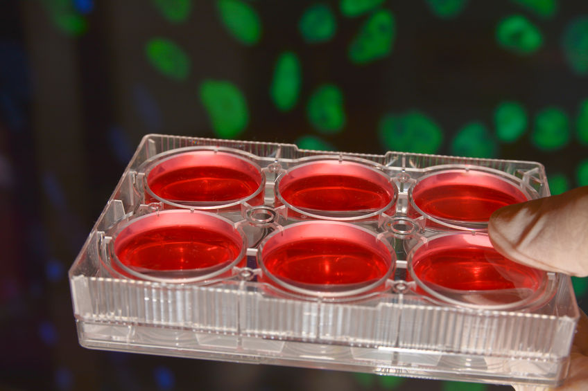
Whenever I write an experiment protocol, among the first things I encounter are calculations regarding necessary cell amount. I have to count the cells prior to the cell culturing procedure or analytical techniques that require an accurate and consistent number of input cells, such as transfection or quantitative PCR. Aiming for an equal number of cells per well is important for standardization of experiments, data integrity, and assay impact—among other things. Additionally, every cell type has different characteristics concerning level of confluence needed before diluting the cells into smaller aliquots for optimal cell growth.
For more than a century, researchers used the hemocytometer to count cells. It’s a manually conducted procedure combined with microscopy. As you can imagine, it is a dreadfully time-consuming and tedious operation. Even among experienced biologists, count-to-count variability was 10-20%. Luckily, we live in the 21st century and most of us are spared from this experience. Nowadays we use automated cell counters, such as Invitrogen Countess II FL Automated Cell Counter. The major advantage is that they provide reliable results in just a fraction of the time required for the traditional way of counting cells. There are, however, a few rules you need to follow and techniques you should master for automated cell counting in order to minimize variability and maximize cell counting confidence.
Sample preparation
The way you handle your samples can improve or diminish counting accuracy and precision. Therefore, a high priority should be put on sample preparation so you can be sure that actual numbers are results caused by an experiment—not by the sample preparation settings. First of all, make sure that you are using standardized procedures when handling cells. I’m referring here to different handling steps, such as proteolysis, pipetting, or centrifugation, which affect total cell count and viability. Here are some pipetting technique tips you may find helpful.
Cell suspensions are disperse systems, and as such have a tendency to precipitate, so make sure that your suspension is homogeneous before mixing it with trypan blue. You can pipette it up and down a couple of times, use gentle vortexing, or manually agitate the tube by hand. Subsequently, pipette the sample from the middle of the tube rather than the bottom, where there is a possibility for a higher concentration of cells.
Principle
Cell membranes are quite selective when it comes to compounds they allow to traverse them, and trypan blue—the diazo dye used in cell counting procedure—is one of the restricted ones. Therefore, live cells won’t get stained, and under the microscope we can easily see the difference between a living cell and a dead cell, which is shown as a distinctive blue color. This principle is also called a dye-exclusion test.
Setting focus
Correct focus setting on your counter is vital for attaining accurate viability information. The live cells need to have a brighter center and slightly darker borders when the focus is set properly. It’s possible that the dead cells look out of focus, but will in any case display a uniform dark staining pattern.
Debris
Depending on the cell type, reagents used, and the experiment protocol itself, debris can be present in different quantities. Debris often originates from contaminated trypan blue, so use it fresh or sterile filter it before use. Debris also comes from dead or dying cells in old cultures, and is generally irregularly shaped, darkly stained, and easily “gated out” using the circularity parameter in automated cell counters.
As with every experimental procedure, cell counting results can be affected by various factors. Sample preparation, focus settings, debris, light intensity, and gating strategies are just some of the few. Luckily, the newest automated cell counters offer optional autofocus, auto bright-field lighting, and multiple gating settings—all of which serve to improve cell count reliability. I can live with these minor glitches, as long as automated cell counting saves me valuable time by avoiding the dreadfulness of manual counting— which, by the way, has even more glitches.
Quartzy is the world’s No. 1 lab management platform. We help scientists easily organize orders, manage inventory, and save money. We’re free and always will be. Visit Quartzy.com or reach out at info@quartzy.com.
Interested in writing for The Q? Send us an email!
Share this:

Tatjana Bosnjak
Tatjana is a PhD Candidate at the Department of Pharmaceutical Biosciences, University of Oslo, Norway. She is interested in how different drugs and substances affect our body, i.e. pharmacology. She studies proteolytic enzymes with a particular focus on cysteine proteases (legumain, cathepsins) and what roles these enzymes have in normal physiology and in diseases such as cancer, atherosclerosis and osteoporosis. Currently, her main goal is to illuminate the cellular and molecular mechanisms of legumain and cysteine cathepsins in bone remodeling and bone cell functions under physiological and pathological conditions of bone loss and to utilize them as potential pharmacological target molecules.