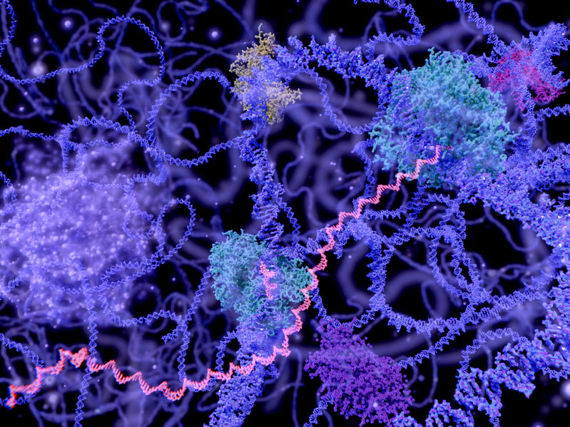
After discussing the importance of DNA methylation, we now examine another functional area of gene regulation: transcription factors, including some of the techniques used to analyze their interactions, and recently published highlights regarding their relevance to pathology. Transcription factors regulate gene transcription and have DNA-binding domains that allow them to bind to enhancer or promoter sequences that can be found at a distance away from the gene to be transcribed. A recent publication (Yin et al., 2017) highlights how specific epigenetic landscaping with CpG methylation affects the binding capacity of transcription factors.
Two of the primary methods used in that paper are SELEX (systematic evolution of ligands by exponential enrichment) and protein-binding microarrays. Using DNA libraries that either had methylated CpG sites or unmethylated CpG sites, they analyzed the ability of different classes of transcription factors to bind. SELEX is powerful because multiple rounds of selection and amplification are used to determine which bind most preferably. What Yin et al. found was that most major classes were inhibited by methylated CpG sites. These are also familiar classes, including bHLH (basic Helix-Loop-Helix), bZIP (basic leucine zipper), and ETS (E26 transformation-specific) transcription factors.
Another method used is the protein-binding microarray to determine the binding ability between transcription factors and DNA. One description discussing the development of this technique is on the website for the Bulyk lab at Harvard, with a link to the 2004 paper. In protein-binding microarrays, there are several distinct ways of detecting bound protein. Medscape by WebMD provides a thorough overview that references “Expert Reviewed Proteomics” of each of the methods, including label-based detection strategies such as fluorescent-tagged oligonucleotides, quantum-dot-conjugated tags, or specifically barcoded, gold nanoprobes. In contrast, label-free methods of detection include the implementation of other familiar techniques, such as detection of the protein-binding complex by mass spec (SELDI-TOF-MS), atomic force microscopy (which can detect topological changes), or surface plasmon resonance, where differences in refractive index are detectable between bound and unbound protein.
So where in the realm of pathology do these findings and methods contribute to the direction of research efforts? An article was recently published on the role of ETS oncogenic transcription factors in tumorigenesis. ETS transcription factors play a role throughout the stages of tumorigenesis, where gene rearrangements, changes in signaling, and mutations at promoter regions lead to changes in ETS activity and, ultimately, tumorigenesis. As a specific example, ETS transcription factor ETV-1 has a role in promoting tumorigenesis in pancreatic cancer and contributes to metastasis and invasion by inducing the epithelial-mesenchymal transition (EMT).
Of course, there are more research areas to discuss beyond cancer. Using protein-binding microarrays as discussed here, a study of families with Mendelian diseases revealed that variants of human transcription factors affected DNA binding affinity, which contributes to the disease phenotype. With the growth of biotech in the area of investigating ancestry and family pedigree, have you checked your transcription factor sets yet?
Share this:

mikecyee
Mike has a Ph.D. in Biomedical Sciences from the University of California, Riverside, a M.S. in Cell and Molecular Biology from San Francisco State University, and a B.A. in English from the University of California, Berkeley.
