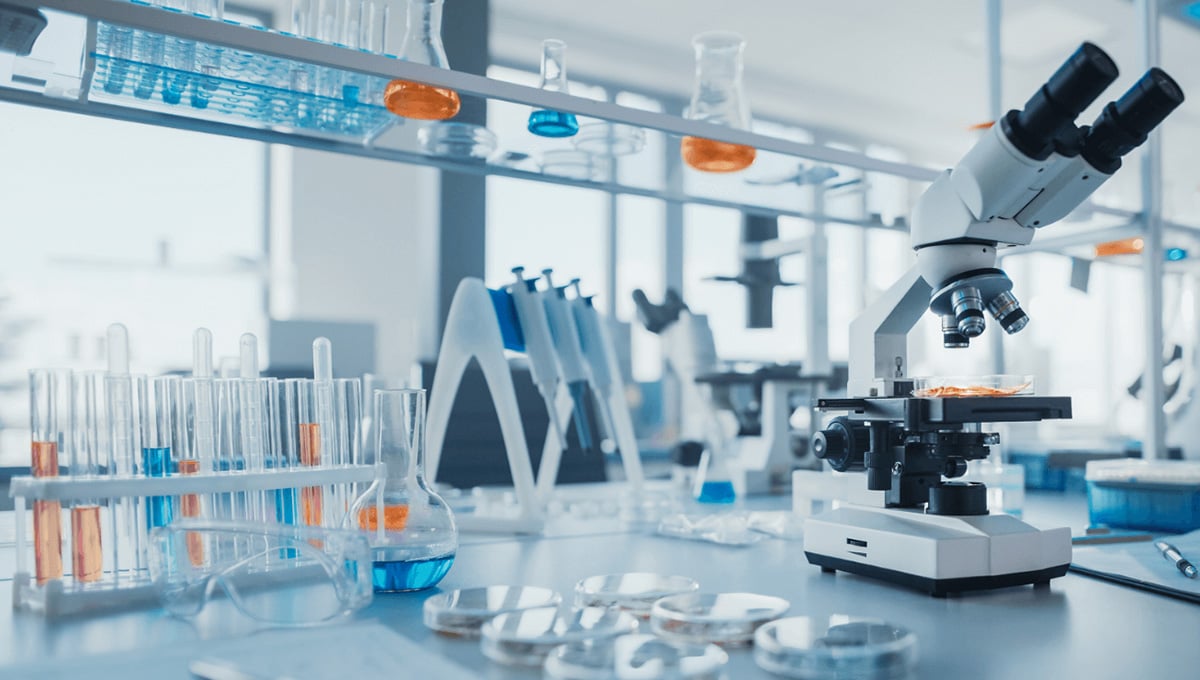
Sterile cell culture practice is key to avoid contamination by microorganisms, which would then interfere with the integrity of your cell system and your experiments. While most of us are careful and well trained in proper cell culture technique, contamination cannot always be avoided. Unfortunately, culture conditions such as the nutrient-rich media and the cell incubator temperature and humidity promote growth of these microbe contaminants and so they grow fast and spread between culture dishes easily. Thus, it is important to be aware of potential contaminants and sources of infection in advance so to be able to best handle them if they do arise.
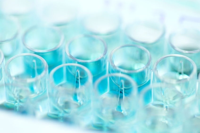
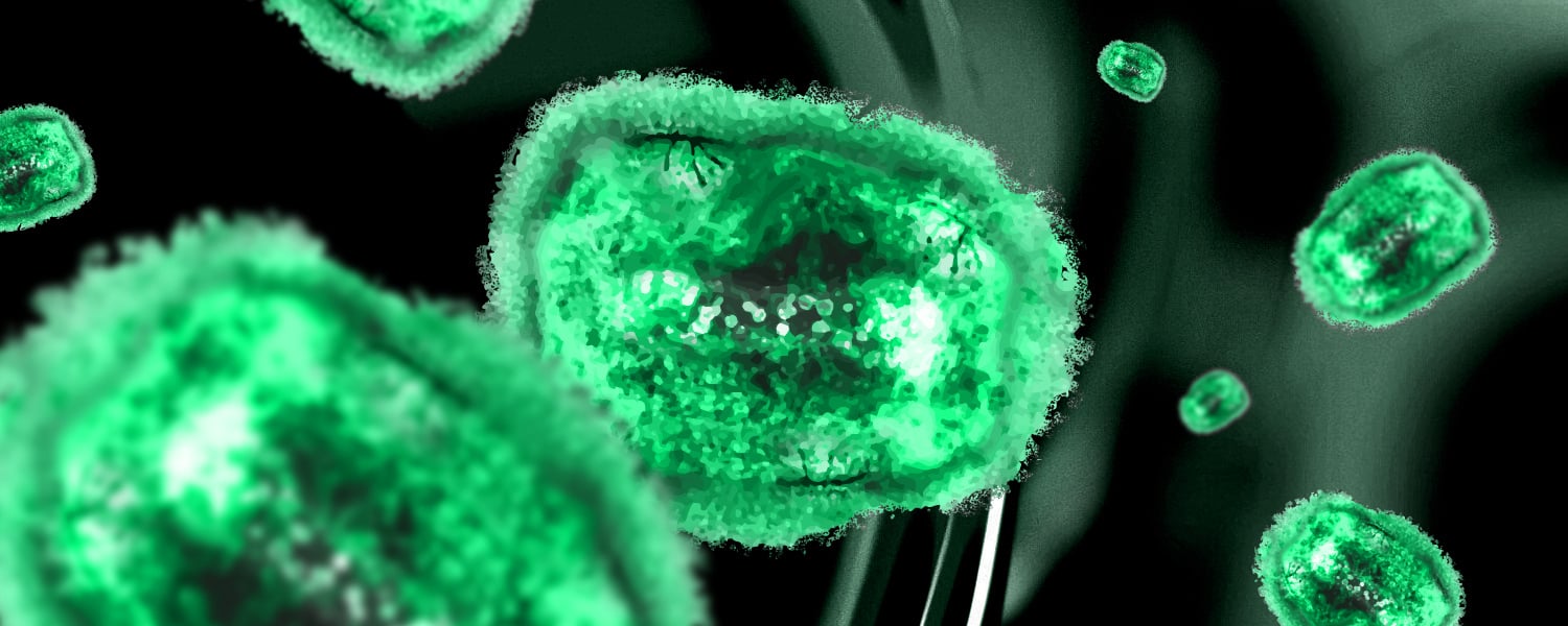
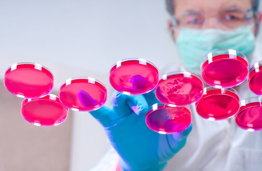
The first step is to know what a contamination looks like. Some contaminants are clearly visible by eye, changing the color and the turbidity of your media. This may look something like when your cells are not attached to the plate but are floating in the media. Other contaminants are not visible by eye, which makes detecting them a challenge. In general, once you do discover that your cells are infected your best bet is to toss them – of course carefully after bleaching the dish they grew in –clean and sterilize the incubator, toss out old media or trypsin that potentially either carried the infection or became infected from your cells, and start fresh. I know this seems extensive, but in the end it will not only be less expensive than having the contamination spread or return and potentially having to re-do your experiments, but also less time consuming. Unfortunately, discarding your cells may not always be an option as, for example, they may be the only copy you have, and so you can attempt to ‘clean’ them. As this may harm your cells, will take time, and keeps the contaminant around longer – potentially to infect other cells - this approach is less favorable. Thus, it is important to be vigilant and to catch infections early to control them. Here are a few main contaminations to watch for.
- Bacteria – Labs try to avoid most bacterial infections through the addition of the antibiotics Penicillin and Streptomycin (Pen/Strep) to the media. However, this is not always sufficient and many labs do not routinely use antibiotics as this could promote antibiotic resistance, or because some antibiotics interfere with cell function. With a bacterial infection you cells will generally look unhealthy, the media may change color or turbidity, and through the microscope you will see moving black dots or rods in the media and the cells. Add a bit of antibiotic to your culture to clean them up. If you are already using Pen/Strep you should add a different antibiotic.
- Fungus – The most common fungal contaminants are mold and yeast. Both are clearly visible in the media, increasing turbidity. Mold will look like long hairy structures under the microscope. Yeast grow rapidly and under the microscope look like clusters with long protrusions sticking out, something like a misshapen pinecone. If either of these infections occur, and you cannot throw out your cells, you have the option of adding an anti-mycotic. These are available from most cell culture companies, often in combination with an antibiotic.
- Mycoplasma – the infection feared the most. Mycoplasma are small bacteria that do not have a cell wall and thus are not affected by treatment with the standard cell culture antibiotics or sterile filtration. Sadly, mycoplasma are practically undetectable without ad hoc testing. There are multiple tests that one can do to check for contamination including PCR, a luciferase assay, or just a quick DNA fluorescent stain of cells. The first two methods are arguably more sensitive and clear cut. By using a DNA stain such as DAPI, you then should visualize extra nuclear spots due to mycoplasma infection. However, from previous experience this method is not as cut and dry, and so it is best to do a longer incubation with the dye or use a higher amount, and to image at a higher illumination intensity to ensure that there is nothing lurking. Unfortunately, mycoplasma often returns to labs. It is hidden in frozen cells or in older reagents or travels from one lab to another. If you are unable to toss your cells for whatever reason, you can clean them with commercially available reagents that seem to be tolerated well by even sensitive cells. With treatment, you will notice that the cells will start to look healthy again and will grow normally.
For precaution, it is advisable to routinely sterilize incubators and to do routine checks. Some labs go further and only make 50mLs of media containing the nutrient rich serum at once and work from that so that if a contamination occurs you would only lose a small amount of media. One way or another, it is advisable that everyone practicing cell culture knows what contaminations to look for and how to handle them to avoid a potential catastrophe.
Quartzy is the world’s No. 1 lab management platform. We help scientists easily organize orders, manage inventory, and save money. We’re free and always will be. Visit Quartzy.com or reach out at info@quartzy.com.
Interested in writing for The Q? Send us an email!